Introduction
According to FIFA statistics, there are about 300 million registered soccer players worldwide, making soccer the most-practiced team sport in the world for males and females of all ages (Dvorak and Junge, 2015). Soccer’s varieties include 11-person classic soccer, indoor and beach soccer and are all performed according to similar rules.
During a match, players do not run constantly as soccer is of an intermittent character (Eniseler et al., 2012). During a professional soccer match, the player's activity includes: walking (41%), standing still (19.5%), running at a low intensity (30%) and running at a high intensity - speed > 5 m·s-1 (8.7%), of which sprinting accounts for only 1.4% (Ferro et al., 2014). Also to be taken into account are running backwards, diagonally, sideways, jumping and landing. The total distance covered by a professional player in a soccer match is between 9 and 14 kilometers (Bradley et al., 2010). Data for the distance and player's activity can differ according to the position on the pitch, anthropometric variables and the performance level. This discipline consists mostly of endurance activity (about 95%), however, its dynamic maneuvers are correlated with success and unfortunately injuries (Chmura et al., 2018; Longo et al., 2012).
The total volume of activities performed during matches and during training often leads to overloads and repeated micro-injuries, and even to acute or chronic trauma. Individual preferences and asymmetrical loading of the lower limbs during kicking and body balancing result in trauma to the thigh muscles, shank and pelvic girdle (Fuller et al., 2006). Fouls also have a significant impact on the injury totals, whose number in the first divisions of England, France, Germany, Italy, and Spain in the 2015/2016 season averaged from 25 to 35 per match (Sapp et al., 2017). Player collisions and overloads are the causes of 1.5–7.6 injuries every 1000 hours of training and 12-35 injuries every 1000 hours of match play on average among elite soccer players (Longo et al., 2012). Moreover, statistical data indicate that youth players are more likely to be injured. As much as 80% of all injuries occur in players under 24 years old, with over 40% cases of those under 15 years of age (Fuller et al., 2016). A low level of agility is a crucial factor predisposing players to injury (Pfirrmann et al., 2016).
Soccer players are most susceptible to hamstring muscle injury, knee ligament rupture, ankle sprain, calf muscle and quadriceps muscle tear, Achilles tendinopathy and groin pain (Ekstrand et al., 2020). It is worth mentioning that many types of injuries, especially ACL injuries, are more likely to affect female players (Montalvo et al., 2019).
Bone fractures or structural bone damages are much less common and comprise only about 10% of all soccer injuries (Robertson et al., 2012). Types of fractures related to soccer are categorized as: acute types, chronic (including stress fractures and avulsion types) and bone cysts. Acute incidences often affect wrists, forearms and tibias (Ekstrand and Torstveit, 2012; Losco et al., 2019; Robertson et al., 2012). Stress fractures are mostly connected with repeated overloading, playing on artificial surfaces, inappropriate adaptation to training stimuli and intensity evidenced by compensations mainly in the form of asymmetrical distribution of muscle forces (Miyamori et al., 2019). Avulsion type bone fractures correlate with age, often occurring in youth players and their immature bones at the site of the tendon attachment. Overloads also have to be taken into consideration (Losco et al., 2019). Other bone structure disturbances such as bone cysts are often discovered incidentally during post injury radiological assessment (Zhang et al., 2020). Bone cysts are painless bone changes that, if left untreated, can lead to complicated fractures (Cho et al., 2019).
Risk factors of soccer player injuries include the player's age, underdeveloped soccer skills, previous injuries, overloads during exercises, volume and intensity of training units, as well as incorrectly conducted treatment and rehabilitation of injuries (Chomiak et al., 2000).
The purpose of this review was to provide a summary of the available research in response to bone injuries in soccer players, simultaneously with the presentation of several uncommon clinical cases. In our present review, we would like to demonstrate the present diagnostic difficulties which complicate correct assessment of the injury, possible treatment procedures and recovery path algorithms. We also wanted to focus attention on motor skills, of which improvement can reduce the risk of various types of injuries.
Stress fractures
Stress fractures are not common, but when they do occur, they are often misdiagnosed in professional soccer, which usually results in a long period of exclusion from training (about 3-5 months). As previously mentioned a young age of a player, as well as an intensive preparatory period may be risk factors predisposing individuals to stress fractures in the extremities (Ekstrand and Torstveit, 2012). Ekstrand and Torstvein (2012) showed that among 2379 soccer players from 2001-2009 there were 51 stress fractures. They were all located in the lower limb, in which the highest percentage were fractures to the fifth metatarsal (MT-5) (78%), tibia (12%) and pelvis (6%).
Stress fractures to MT-5 versus acute fractures (arising from the mechanical trauma: kick or strike) are much more common and account for nearly 80% of all cases (Miyamori et al., 2019). Injury and accompanying pain may occur when loading the lateral edge of the foot in an uncontrolled position, which often occurs during physical activity, such as running, playing soccer, playing basketball, baseball, etc. (Kuzuyama et al., 2019; Singh et al., 2018). Researchers have shown several important factors which increase the risk of this fracture. Raikin et al. (2008) reported that many patients with MT-5 fractures have evidence of varus hindfoot alignment. Saita et al. (2018) noted that increased risk of fractures occurred in people with restricted internal hip rotation. Miyamori et al. (2019) showed that a field surface can have a significant impact on MT-5. Soccer players who spent more than 80% of the play time on artificial turf pitches were 2.5 times more likely to have MT-5 fractures than players playing on natural grass. The same study also indicates that an MT-5 fracture is more likely to affect the non-dominant leg.
The treatment depends on the severity of the injury. In the treatment of minor fractures, in which bone migration has not occurred, it is sufficient not to load the injured limb and to use footwear with a hard sole. Temporary immobilization of the limb is also sometimes recommended. The typical length of immobilization is from 6 to 10 weeks, and healing time is typically up to 12 weeks. For serious injuries, delayed union and non-union injuries surgical intervention is advised, in the form of internal fixation, to facilitate bone connection. In addition, removal of bone fragments is sometimes necessary. A plantar gap greater than 1 mm is predictive of a prolonged bone union and bone grafting can be helpful in this case. The average recovery time after intramedullary MT-5 immobilization is about 9 weeks (Pecina et al., 2011; Miller et al., 2019). It has been shown that return to play after fewer than 8 weeks results in a higher risk of delayed radiological union. However, this fact neither prevented the athlete from continuing to play soccer nor did it ultimately affect the risk of non-union (Miller et al., 2019). For conservative treatment the major benefit is avoiding surgery, while the main risk is the development of non-union. For surgical intervention, the main benefit is an improved return rate and time to sport, while the main risk is connected with such undesired side effects as structural damage and infection (Robertson and Wood, 2017).
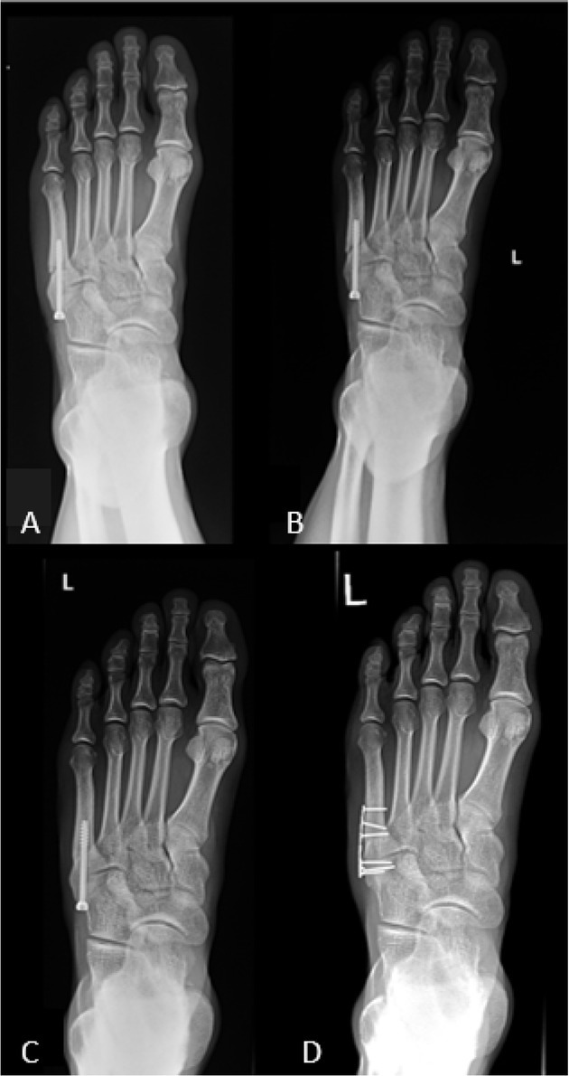
Among more interesting cases of stress fractures there was a right-footed 27-year-old professional soccer player, injured during a match, whose treatment combined surgical intervention, orthobiologic treatment and rehabilitation. X-ray imaging showed a fracture at the base of MT-5 in the left foot. The decision was made to surgically repair the fracture using a cannula screw (4.5 mm/45 mm) (Figure 1A, 1B). After the procedure, a Walker brace boot was recommended along with crutches. The patient did not report any pain. The motor control improvement process was recommended after 14 days, followed 6 weeks later by running on soft surfaces. After 9 weeks, during the follow-up visit, platelet rich plasma (PRP) (2 ml) was applied to enhance the quality of the bone union. After return to play nearly three months after the injury, the patient suffered another injury due to being kicked in the operated foot by an opponent. X-ray images showed a partial fracture of the left MT-5 (Figure 1C). An additional surgery was not necessary in this case. A 2-week break from soccer training was recommended. During the control visits (one and two weeks after the fracture) PRP (2 ml) was administered. Eight weeks after the second injury, the patient still reported pain during training. It was decided to surgically replace the screw with a Hofer plate (Figure 1D). The patient experienced pain for 4 weeks after the surgical procedure. After 8 weeks, 2 ml of PRP was applied to the patient and further rehabilitation was recommended. Ten weeks after the second fracture he was allowed to return to full sport activity.
Figure 1
X-ray of metatarsal fractures five: A – direct after the first surgery (intramedullary screw fixation); B– one month after the first surgery; C– after the second injury; D– after the second surgery (intramedullary screw was changed to a tubular plate).
First signs and symptoms of a tibial stress fracture generally include pain and tenderness on the medial shaft of the tibia which increase with exercise. Usually training is possible, however, with discomfort. Much like in the other types of stress fractures, plain radiographs have a high false-negative rate, and MRI or CT are more sensitive and precise types of diagnostic imaging (Robertson and Wood, 2015). In the early stages, only one tibia may be affected, but later changes can be seen in both legs (Gmachowska et al., 2018). A plethora of therapies are taken into consideration, but the most reliable treatment of the fracture is ensured by orthopaedic surgery. A conservative modality of treatment using even orthobiologic therapy (autologous blood, PRP, autologous conditioned serum and stem cells), usually extends the time needed to return to play compared to surgical treatment. It was found that the use of a nail or plate reduced time to return to sport, with an acceptable complication profile (Robertson and Wood, 2015).
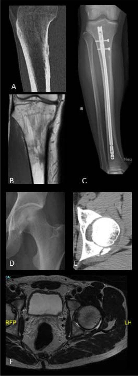
One example of surgical treatment for a tibial stress fracture involved a right-footed 21-year old professional striker. He complained of pain in the right tibia area which lasted for 14 days and intensified during physical activity. Medical examination revealed tenderness and a widening contour of the 1/3 proximal right tibia zone. Normal knee and ankle mobility and stability were confirmed. MRI in correlation with CT and ultrasound confirmed a chronic stress fracture of the right tibia at the site adjacent to the popliteus and soleus muscles attachment (Figures 2A, 2B). Due to the extent of the damage and the requirements of his sports activity, it was decided that a surgery union with a titanium intramedullary nail would be performed (Figure 2C). For 2 weeks after the procedure, orthosis and elbow crutches were recommended. The rehabilitation process was initiated as soon as possible. Isometric exercises were recommended as part of rehabilitation, and stabilization exercises were introduced 2 weeks after the surgery. The patient started training after 4 weeks and after 2 months he was allowed to return to full sport activity.
Figure 2
Stress fracture: A - the tibial before the surgery (TK); B- the tibial before the surgery (MRI); C- the tibial fixed intramedullary nail (X-ray); D- the iliac acetabulum (X-ray); E- the iliac acetabulum (CT); F- the iliac acetabulum 6 months after the injury (MRI).
Pelvis stress fractures are very difficult to diagnose, because pain associated with these injuries is a fairly common symptom in various types of deteriorations in the pelvic area. Due to the spatial anatomical proximity of the groin to the hip, pubic bone, fascia, muscles and nerve structures of the abdominal wall, pain can have various causes, posing significant diagnostic difficulties (Hölmich et al., 2004). It is estimated that groin injuries affect 4–19% of all injuries in men and 2–14% of all injuries occurring in women (Weir et al., 2015). In the pelvis area there are several sites predisposed to a stress fracture: sacral bone, pubic bone and the pubic rami (Tins et al., 2015). Femoroacetabular impingement (FAI), which is very common, hinders the diagnosis of the fracture in the groin region. Due to the similarity of symptoms caused by tissue overuse and fracture, playing soccer through the injury often worsens the local tissues. The causes of this type of injury are usually an accumulation of micro-injuries, which in further stages, can lead to acetabular labrum damage. This injury can occur unilaterally or less often bilaterally, most often affecting people in their 30’s or 40’s (Ganz et al., 2003). The treatment approach for the pelvis stress fracture is similar to other stress fracture cases. In cases characterized by a very high probability of bone union, conservative treatment is usually enough (Tins et al., 2015).
Such treatment was adopted for a right-footed 53-year-old amateur soccer player diagnosed with a fracture of the iliac acetabulum. During a soccer match he felt a stabbing pain in his left hip as a result of a sudden stop. After the incident, the patient felt pain while performing rotational movements. The source of pain was located in the trochanter area and the medial side of the hip. The X-ray image showed a bone thinning in the acetabulum of the left hip (Figure 2D). Further diagnosis was recommended using CT, which revealed a fracture of the posterior iliac acetabular with displacement of bone fragments (Figure 2E). In addition, a slight indentation fracture of the femoral head in the pit area was observed, with the presence of small bone fragments. Conservative treatment, supported by myofascial therapy of the hip and thigh area, was applied. Dynamic loading and rotational movements in the hip joint were introduced. After a period of 2 months from the occurrence of the fracture, PRP (2 ml) was administered to the left hip joint from the trochanter side. The myofascial therapy in the hip and thigh area, torso stabilization and rolling were recommended in the rehabilitation protocol. For 5 months dynamic loads and rotational movements in the hip joint were to be avoided. Approximately 5 months after the injury, the patient regained the ability of independent but limping gait. Control MRI was performed after 6 months (Figure 2F) and further rehabilitation was recommended up to 11 months after the incident, leading to recovery of left hip stability, with no pain reported during daily activities. Abstaining from soccer play was recommended.
Avulsion fractures
Avulsion fractures occur when muscle strength exceeds mechanical properties of bones. This type of fractures are most often observed in young, skeletal immature athletes resulting from dynamic and explosive types of motion (Losco et al., 2019). In mature athletes, avulsion fractures are very rare and their cause is usually microtrauma or less often direct trauma (Khemka et al., 2014). Diagnostic imaging must overcome the challenge of distinguishing the growth plate in non-adults from the real fracture.
Complementary diagnostic imaging protocols are suggested in the form of MRI and CT scans (Losco et al., 2019). Avulsion fractures are rarely suspected as a cause of groin pain (Otto et al., 2020). Eberbach et al. (2017) showed the frequency of various hip avulsion sites as follows: the anterior inferior iliac spine at 33.2%, ischial tuberosity at 29.7%, anterior superior iliac spine at 27.9%, iliac crest at 6.7%, lesser trochanter at 1.8%, and superior corner of the pubic symphysis at 1.2% of all cases. Ball game disciplines were the primary predisposing factor of the two first cases (70% and 45%, respectively).
Avulsion fractures of trochanter minor are very rare injuries described in only few publications, representing <1% of all hip injuries (Dukas et al., 2019). All cases described in the literature arose from a sports context and occurred in adolescents (13.7 years old; range from 9 to 17) (Memminger, 2018).
The avulsion fracture of the ischial tuberosity is the next most common cause of hip related injuries, occurring mainly among younger players (average age is 13-17 years old). Susceptibility to these injuries is associated with ischial tuberosity ossification. This process begins between the age of 13 and 15 years old and finishes between 20 and 25 years old. During this period the elasticity of the epiphysis is 2 to 5 times weaker, which makes it easier to separate the epiphyses from the tarsal plate. Ischial tuberosity avulsion fractures can cause significant diagnostic difficulties and are oftentimes classified as a proximal hamstring injury, intervertebral disc disease, piriformis syndrome or ischial tuberosity bursitis. Sometimes late diagnosed avulsion fractures are similar to a bone tumor (osteosarcoma or osteochondroma) (Liu et al., 2018).
There was no significant advantage of any treatment method. As opposed to the management of sports stress fractures, for avulsion fractures usually conservative treatment is suggested, except for incidents when a retraction of the separated bone insertion may reduce the efficacy of the bone-tendon unit, not to mention other potential complications such as non-union heterotopic ossification, muscle shortening and fibrosis at the muscle origin (Elattar et al., 2016). Surgical treatment is considered the treatment of choice for fractures with a displacement of more than 15-20 mm. Additionally, it was demonstrated that surgical treatment led to faster implementation of rehabilitation procedures and an earlier return to full activity (Eberbach et al., 2017; Khemka et al., 2014). Conservative treatment consists of partial weight bearing on the limb using crutches and taking NSAIDs from 3 to 6 weeks. The return to sports activity is possible after an average of 2-6 months. In surgical procedures, a full load is allowed after 4.5 weeks on average and returning to sport after 2.4 months (Eberbach et al., 2017).
One example of a conservative treatment used for an avulsion fracture involved a 28-year-old patient who complained of severe pain in the hip area after a collision with an opposing player during a soccer match. A separated bone fragment (length 26 mm, width 39 mm) of the lateral iliac bone above acetabulum in the gluteus minimus muscle belly area was visible in the pelvis x-ray image (Figure 3A) and in the ultrasound. Six millimeters bone plate dislocation was confirmed based on MRI imaging (Figure 3B). In addition, this area and of the surrounding soft tissues displayed an increased signal as in the case of edema. After consultation with a radiologist, it was decided the patient should be treated conservatively. Physiotherapeutic treatment, with functional exercises emphasizing core stability, was recommended. Exclusion from full training loads took 40 days.
Figure 3
Avulsion fracture: A - X-ray of the left lateral iliac bone; B- MRI of the left lateral iliac bone; C- X-ray of the right ischial tuberosity before the surgery; D- X-ray of the right ischial tuberosity after internal fixation;
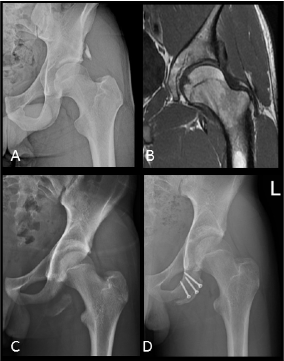
A different approach was adopted for a 22-year-old athlete who felt severe pain around the right buttock during soccer training (while running at high intensity). Extreme tenderness was noted during palpation of the ischial tuberosity area. Pain arose during knee flexion. Based on ultrasound imaging, swelling around the sciatic tumor and the hamstring muscle group attachment was observed. X-rays confirmed the avulsion fracture of the right ischial tuberosity (without significant displacement) and rupture of the muscle attachment. The patient was qualified for reconstruction of the proximal hamstring muscle group attachment. Resistance exercises with a slight load after surgery and increases in their intensity 3 months after were suggested. Four months after the surgery the patient did not report pain and completed the treatment. In very serious cases, where the bone is displaced, broken off and/or significantly dislocated, screws should be used for fixation. This serious case is shown in Figures 3C and 3D.
Bone cyst
A simple bone cyst is a space devoid of proper bone tissue, which in the majority of cases is a benign variety of a bone tumor. In X-rays, they are visible as oval thinning of bone tissue. Bone cysts are most commonly located at the epiphyseal of the humerus, femur, and tibia. Most of them are detected incidentally as a bone structure disorder during the diagnosis of a different injury (Zhang et al., 2020). The most probable etiology is related to the blood supply and alterations of the fluid pressure inside the cyst space. The result of increased pressure in the bone is an increase of inflammatory protein markers released by endothelial cells, causing osteoclastic activation and bone resorption (Savic et al., 2019). To avoid a fracture at the site of the cyst location, it is recommended that large bone cysts be treated surgically by filling them with bone transfer or with the biomaterial agents (Cho et al., 2019). Only small cysts may be treated using corticosteroid injections. Treatment of bone cysts usually lasts several weeks and is a kind of a preventive therapy to avoid a fracture at this site. A quick return to full fitness and physical activity is possible. Due to the unknown etiopathogenesis etiology of the lesion, histopathological examination is advisable (van de Luijtgaarden et al., 2009).
One interesting case involved a bone cyst located in calcaneus. A 15-year-old soccer player complained of heel pain, especially during intense training and playing on artificial turf. X-ray examination showed a spherical radiance in the anterior part of the right calcaneus 1.5 cm in a diameter (Figure 4A). To advance the diagnosis, MRI was used, indicating the presence of a single bone cyst (13 x 14 x 24 mm) (Figure 4B). The method used was to fill the cyst with allogeneic bone and for the limb to remain in a non-weight-bearing state for one week. The patient returned to play after 10 weeks.
Figure 4
Bone cyst: A – X-ray of the cyst in heel bone; B– MRI of the cyst in heel bone; C– X-ray of a distal segment of the right humeral bone; D– X-ray of the humerus cyst filled with bioresorbable material; E– X-ray of the proximal humerus cyst; F– X-ray of the proximal humerus cyst, one year after methylprednisolone was applied.
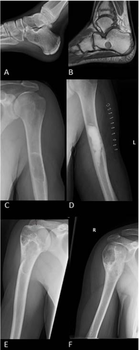
Another case of a bone cyst, which was detected before the fracture occurred, appeared in a 28-year-old player who felt pain in his left arm for 3 weeks. The patient negated the traumatic background and reported increased pain during palpation. A simple cyst at 1/3 of the distal humerus was visible in the X-ray image (Figure 4C). In the MRI, at 1/3 of the distal and medial humerus, fluid signal changes were indicated. The cyst was surgically removed and the gap filled with bone substitute material (Cerament) at 18 ml volume (Figure 4D). Immobilization of the operated limb until the date of the control visit (14 days after surgery) was recommended. The patient did not report any pain or side effects. A two week break in training was recommended.
Another case of a different character involved a 16-year-old soccer player who visited the clinic after a humeral bone fracture. X-rays performed after the injury showed, apart from the fracture, the presence of a single bone cyst at the fracture site (Figure 4E). As a result of the weakened bone structure due to the cyst even normal loads caused the fracture. Initially, steps were taken to enable bone union, followed by treatment of cysts (8 months after the fracture). For the treatment of cysts, an endosteal injection of methylprednisolone 40 mg/ml was used and a sling was recommended for 4 weeks. After 5 months, a second dose of methylprednisolone was given. After 6 months of healing, about 50% of the cyst was observed (Figure 4F) and recreational physical activity was allowed. After one year from the first administration of the preparation, he was allowed to return to professional sports activity.
Prevention
One of the ways to reduce the number of injuries in sport is to incorporate improvement exercises into the training sessions with the aim of injury prevention. An important step in this field was the introduction of the FIFA 11+ program, of which positive impact on preventing injuries has been reported in many publications (Sadigursky et al., 2017; Silvers-Granelli et al., 2015). Interesting solutions were also put forward by the Swiss National Insurance Fund, the Suva "Sport Basics" (SSB) program, of which the theme was injury prevention for all ball sports (Gebert et al., 2019).
The key points of improvement programs seem to be an adequate level of agility and motor control to safely assign training loads in an adaptive manner and avoid improper tissue compensation. Statistics show a clear reduction in the risk of soft tissue injuries (mainly in the knee area) against biomechanical factors in athletes following neuromuscular training (Donnell-Fink et al., 2015; Hewett et al., 2006; Stastny et al., 2015). In addition to preventive properties, it has been found that performing exercises from the FIFA11+ protocol improves agility, balance, jump height and muscle strength (Trajković et al., 2020).
It should be emphasized that better overall body coordination and stability contribute to injury prevention (Gatterer et al., 2018). This thesis is confirmed by a higher injury rate in young players, compared to professional adults whose training experience and proficiency level are higher (Pfirrmann et al., 2016). The susceptibility to injury increases with player’s fatigue, as evidenced by a higher ratio of injuries sustained during the match than during training (Longo, 2012). The reason for this phenomenon may be a lack of knowledge or poor physical preparation, as well as the fact that decidedly more people do sport recreationally than competitively (Berczyński et al., 2017).
Summary
Bone injuries represent a small percentage of all soccer injuries, but they can exclude a player from the game for a long time. The occurrence of the bone deteriorations may have relevant health implications and impact on the athlete’s career. The cases presented in this manuscript are special types of bone injury of which a diagnostic process is more problematic than their treatment. For prevention it is recommended to play on natural grass and to use appropriate footwear, which, if necessary, should be equipped with orthopedic insoles. Adequate physical and motor preparation are even more important, from the standpoint of injury prevention. Adequate and systematic recovery protocols combined with physiotherapy will certainly contribute to reducing the rate of reinjury. It is important to note that X-ray and ultrasound, as the primary diagnostic tools, can be inadequate for some types of fractures. With persisting symptoms and no clear radiographic changes, more precise tools such as MRI and CT should be involved. The treatment of fractures is an individual issue and depends on the type of damage. To accelerate the return to play, especially for professional athletes, surgical procedures are recommended. To enhance the quality of the healing, some biological factors as autologous blood, PRP, autologous conditioned serum or stem cells may be applied.
In order to optimize the return to sports activity, even at the performance level before the fracture, it is necessary to regain the highest quality of bone tissue as well as adequate levels of fitness necessary for the sports discipline.