Introduction
Specialised indoor bicycles manufactured using a weighted flywheel at the front are used in indoor cycling exercises called spinning. Cycling indoors using an immobile spin bike has gained popularity as a training modality for triathletes. Part of its appeal is that indoor spin cycling can form part of a structured and periodised training program and provide an alternative to outdoor cycling when faced with environmental extremes of cold, hot and/or hypoxic environments. In this regard, indoor spin cycling allows triathletes to preserve cardiovascular fitness levels and training loads.
In cycling, the lower extremities are responsible for producing energy imparted to the bicycle. To mediate this effect, triathletes continuously adjust the force and timing produced relative to the pedal position to obtain a specific self-selected pace (de Melo Dos Santos et al., 2017). A change in the body position may alter cycling variability due to both fatigue and mechanical factors (Padulo et al., 2012).
A characteristic of efficient movement control in cycling is that of trunk stability. Cycling requires trunk stabilisation in order to balance the bicycle (McDaniel et al., 2005). A forward shift of the whole body centre of mass (CoM) means that the cyclist’s body is less supported by the saddle and would require additional stabilisation from the trunk. Similarly, a change in the cycling position when moving from the drop handlebars to an aerodynamic position would alter the whole body CoM. A low level of core trunk muscle strength or stability can cause additional upper body movement. In this instance, the capacity of the lumbar-pelvic-hip complex to control trunk movement and preserve trunk stability can be compromised. Therefore, the core musculature may influence the kinematics and load-bearing capacity of the knee by determining what loads are transmitted from the trunk. Taken together, poor trunk strength or stability combined with increases in power output could cause excessive CoM acceleration leading to inefficient movement. The idea being that higher intensity levels are related with larger mediolateral force swaying (Rannama et al., 2017) with strenuous cycling decreasing stability in the anteroposterior direction (Wiest et al., 2011). Correspondingly, an increase in the workload necessitates additional upper body stabilisation (McDaniel et al., 2005).
During incremental, non-steady state exercise, a point is reached at which a participant’s ventilation shows a non-linear increase, which is termed the ventilatory threshold (Ghosh, 2004). Relative to cycling, this threshold implies an effort that exceeds the steady state. In steady state cycling (i.e., changes to intensity vary only negligibly over a specified time), the forces involved in pedalling largely fluctuate throughout the crank cycle (Gregor, 2000). While these designs have been utilised to inform decision-making as to the effects of indoor cycling, studies involving steady state indoor cycling are limited in comparison.
A multi-method approach, including intensity measured as relative (i.e., as a percentage of maximal oxygen uptake, VO₂max) or absolute (as metabolic equivalent of tasks, METs), where one MET is defined as the resting metabolic rate (RMR), can be employed. Yet, this presents limitations as without direct measurements, the RMR is usually replaced by a 1-MET reference value of 3.5 ml O₂kg1min1(Ainsworth et al., 2011). Absolute and relative energy expenditure – EE (kcal, kJ, MET) can be estimated by caloric cost. Although notwithstanding its own limitations, the MET is a method to indicate and compare the absolute intensity and energy expenditure of different physical activities (American College of Sports Medicine, 2008); thus, the MET is a measurement of exertion intensity (Pollock et al., 1998) and has been used alongside the rating of perceived exertion (RPE) during graded cycle ergometer tests (Kasimary Çakir et al., 2012). The MET and RPE may be attractive due to their noninvasive nature, low cost, squad-wide concurrent measurement capability and time efficiency.
The relationship between the RPE and physiological markers such as the heart rate (HR) has been strongly correlated (Scherr et al., 2013) and often demonstrated during passive estimation and perceptually regulated procedures (Garcin et al., 1998). Measurement of the RPE remains of interest because the scale also predicts oxygen uptake (VO₂) (Wong et al., 2011). In cycling, the RPE is usually applied either as a means of estimating pre-selected power output or producing appropriate power output for a preselected RPE (Myles and Maclean, 1987). This method assumes that participants can adjust power output to match numerically anchored verbal expressions of effort.
The use of the RPE in the prescription of alternated power protocols during indoor spin cycling assumes a comparable relationship between indices of physiological mechanisms inclusive of the heart rate and heart rate reserve (HRR) usually measured during continuous laboratory tests. These relationships appear to follow identical trends for continuous and intermittent exercises with the same power output (Edwards et al., 1972). Yet, the influence of whole body CoM acceleration on these variables remains largely unexplored in spin cycling, particularly in the triathlete population.
An objective in cycling is to eliminate all unwanted postural movement and reduce unused force. This unwanted force measurement is possible using accelerometers. Advances in wearable technology have made the accelerometer the device of choice for measuring frequency, duration and intensity of activity (Sallis and Saelens, 2000) as they provide an objective assessment of energy expenditure (Crouter et al., 2018). The accelerometer presents as a tool to provide evidence for coaches and triathletes to monitor the trunk position via accelerations to the whole body CoM whilst performing varied spin cycling protocols during training and competition. Consequently, quantification of CoM acceleration can be detected. Therefore, the aims of this pilot study were to (1) identify any differences between two commonly used triathlete bicycle racing positions (aerodynamic and drops), which may cause variations to the whole body CoM; and (2) investigate the association between the whole body CoM, RPE, MET and HRR during steady state indoor spin cycling. Quantifying these variables could provide a better understanding of the functional significance of the whole body CoM in order for new insights into postural control mechanisms.
Methods
This observational pilot study recorded accelerations to the whole body CoM in recreational triathletes during a characteristic spin cycling training session which was conducted in the triathlete’s natural training environment.
Participants
Four triathletes (3 males, 1 female) (mean ± standard deviation [SD]; 31 ± 8.8 years, 77 ± 8.5 kg, 178 ± 0.59 cm, 8.7 ± 3.71 weekly training hours) were recruited through contact with the local triathlon club (Greensborough Triathlon Club). All participants were asymptomatic of illness and free from any acute or chronic injury, as established by the American College of Sports Medicine (2008) participant activity readiness questionnaire (PAR-Q). To be included participants had to be cycling at least 50 km a week and competing in triathlon for a minimum of 12 months. Participants were excluded if they were sensitive to double sided tape or had an injury that might affect their riding kinematics. Written informed consent was provided, which was approved by Charles Darwin University Ethics Committee (HREC19028).
Procedures
The magnitude of whole body CoM acceleration was continuously measured by a single triaxial accelerometer (52 x 30 x 12 mm, mass 23 g; resolution 16-bit, full-scale range 16 g, sampling at 100 Hz: SABEL Labs, Darwin, Australia) (James et al., 2011) which was attached between the L5 and S1 spinous process (Kavanagh et al., 2006) using double sided elastic adhesive tape (Medtronic Australasia Pty Ltd, Macquarie, NSW). This position was selected as it is the closest external point to the whole body CoM and the lowest point where a single measuring device can monitor left and right lower limb kinematic data (Winter et al., 2016). Each participant performed one indoor 20 min cycle which consisted of six different power output conditions (Watts) and two body positions (DP = drops, AO = aerodynamic) (Table 1).
Table 1
Protocol to assess body CoM acceleration, RPE, MET, HRR at 6 spinning conditions. FCPO = freely chosen power output; DP = drops position; AO = aerodynamic position
| Epoch (minutes) | 2-5 min | 5-8 min | 8-11 min | 11-14 min | 14-17 min | 17-20 min |
|---|---|---|---|---|---|---|
| Power (Watts) | FCPO | <150 | >152-205 | >152-205 | >206-246 | FCPO |
| Position | DP | AO | AO | AO | AO | DP |
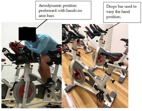
The DP position is described as hands placed onto the bicycle drop bars with slight elbow flexion and trunk inclination. The AO position is described as using tri (aerodynamic) – bars with elbows on triathlon bars with a greater horizontal torso than the DP position (Figure 1).
Figure 1
(a) Participant cycling in the aerodynamic position; (b) Schwinn Carbon bicycle used in the study
Participants completed the cycling conditions at their customary training location. The room temperature was standardised and controlled at 19°C using a climate control system. Cycling was conducted in the evening with participants cooled with two ventilated fans and permitted 250 ml of water. For the bicycle set up, participants were asked to configure the handlebar and saddle position to their preferred settings on a Schwinn Carbon Blue bicycle (Schwinn Bicycles, Dorel Industries, Inc, Washington, USA) which consisted of a belt drive (122 x 109 x 51 cm). Strap-in pedals were used to replicate a typical training setting in order to limit potential confounding effects. Power output was displayed via the digital display unit on the bicycle which was maintained using visual feedback. When a change in power was required (i.e. at three minute epochs), participants were verbally instructed to adjust gear ratios that would sustain the level of intensity required. To ensure familiarly, power output was based on a typical training session. Participants were required to refrain from any strenuous exercise for 24 hours before testing. Prior to commencement, participants performed a self-selected 2 min warm-up at their freely chosen power output. Participants were asked to verbally signify exertion using the Borg 6-20 RPE scale (Borg, 1998) at the end of each epoch. Heart rate reserve (HRR) was estimated by extrapolation of the RPE by 10 to obtain individual beats per minutes (Borg, 1974). All participants had previous experience of RPE perceptual scaling. The accelerometer was controlled wirelessly by the principal author via a standard Hewlett Packard PC using a comprehensive MATLAB Toolkit. This allowed for control of multiple accelerometers providing no restrictions during data capture. Data were subsequently downloaded from the accelerometers using a SABEL Sense software program (SABEL Sense 1.2_x64, SABEL Labs) via a CSV file. Prior to cycling, the accelerometer was calibrated (Lee et al., 2018). The accelerometer was synchronised at the start and the end of each three minute epoch where the longitudinal (vertical), mediolateral and anteroposterior axis aligned with X, Y and Z, respectively. Data were recorded continuously.
Data analysis
Cycling conditions and corresponding power changes were identified in the raw accelerometer data. Longitudinal acceleration (CoMLN = sqrt (X2) was used to identify a change in posture and was identified when the acceleration magnitude began increasing towards its largest peak. Mediolateral acceleration (CoMML = sqrt (Y2)) was used to identify pedal strokes. For each epoch, whole body CoM acceleration magnitude (CoMtotal = sqrt (X2+ Y2+Z2)) was then hand scored to obtain a 60 s average in order to attain a true reflection of steady state cycling. To obtain the MET compendium codes to be included in our study, researchers from this paper reviewed the 2011 Compendium of Physical activities (Ainsworth et al., 2011) to select the description which best matched the activities. The MET values were subsequently classified as: (1) conditioning exercise bicycling, stationary, 8.8 MET (101–160 W, vigorous effort); (2) conditioning exercise bicycling, stationary, 11 MET (161–200 W, vigorous effort); (3) conditioning exercise bicycling, stationary, 14.0 MET (201–270 W, very vigorous effort). To convert mph to kilometres per hour (km/h) a conversion factor of 1.609344 was applied to ensure consistency with the digital display screen on the Schwinn bicycle. To obtain a 60 s average, the caloric cost was calculated according to Equation 1:
Equation 1
where MET is multiplied by oxygen uptake (VO₂) of approximately 3.5 O₂kg1min⁻1ml/kg/min⁻1multiplied by weight in kilograms.
Statistical Analysis
Prior to any inferential statistical analyses, descriptive statistics were checked for normality using quantile–quantile plots. Statistical analysis was completed using Analyse-it (Leeds, United Kingdom, version 4.92). Results correspond to the mean ± SD. The analysed variables were: (1) mean value of whole body CoM acceleration magnitude; (2) mean value of the MET and caloric cost; (3) mean value of the RPE; and (4) the contribution of both drops and aerodynamic position to all analysed variables. Values were tested using 95% confidence intervals (CI) and threshold values classified as 0.1–0.3 (small), >0.3– 0.5 (moderate), >0.5–0.7 (large), >0.7–0.9 (very large) and >0.9 (extremely large) (Hopkins et al., 2009). A paired-sample t test assessed differences in variables with an alpha (α) level set at 5% (p < 0.05). Two SD represents the expected change in whole body CoM acceleration given a 2 ± SD change in power, or otherwise the change from a typically low (−1 SD). The Pearson’s product-moment correlation coefficient (r) measured the strength of the relationship between the dependent variable of the MET and both the whole body CoM acceleration and distinct magnitudes (i.e., longitudinal, CoMLN) along with the RPE and HRR.
Results
All participants completed the six cycling conditions. Effect sizes revealed mostly small differences in both longitudinal and anteroposterior acceleration (< 0.2) throughout all conditions. The exception was found from epoch 8–11 min to 11–14 min performed at 152–205 W with extremely large effects (> 0.9) for all acceleration magnitudes as well as the RPE and HRR, the latter with greater variability given the disparity in standard deviation (Table 2).
Table 2
Mean ± SD and effect size for CoM acceleration magnitude (m/s2), RPE, MET and HRR, stratified by epoch and condition
| **Epoch 2-5 min (FCPO) | *Epoch 5-8 min <150 w | Threshold Effect Size (ES) | |||
|---|---|---|---|---|---|
| Direction | Mean | ± SD | Mean | ± SD | Descriptor |
| X | 25.19 | 3.28 | 24.50 | 3.31 | -0.2 (small) |
| Y | 1.35 | 1.36 | 0.74 | 0.62 | -0.5 (moderate) |
| Z | 12.83 | 6.39 | 12.06 | 5.51 | -0.1 (small) |
| RPE | 6.75 | 0.50 | 11.75 | 1.89 | >0.9 (extremely large) |
| HRR (bpm) | 67.5 | 5.00 | 95.00 | 17.32 | |
| MET | 6.00 | 6.00 | |||
| **Epoch 5-8 mins <150 w | *Epoch 8-11 mins 152-205 w | ||||
| X | 24.50 | 3.31 | 24.74 | 4.10 | <0.1 (small) |
| Y | 0.74 | 0.62 | 0.77 | 0.59 | <0.1 (small) |
| Z | 12.06 | 5.51 | 14.34 | 8.46 | <0.1 (small) |
| RPE | 11.75 | 1.89 | 11.75 | 1.89 | <0.1 (small) |
| HRR (bpm) | 95.00 | 17.32 | 117.5 | 18.93 | |
| MET | 6.00 | 8.00 | |||
| *Epoch 8-11 mins 152-205 w | *Epoch 11-14 mins 152-205 w | ||||
| X | 24.74 | 4.10 | 24.90 | 4.07 | >0.9 (extremely large) |
| Y | 0.77 | 0.59 | 1.24 | 1.05 | >0.9 (extremely large) |
| Z | 14.34 | 8.46 | 14.39 | 8.17 | >0.9 (extremely large) |
| RPE | 11.75 | 1.89 | 12.25 | 1.50 | >0.9 (extremely large) |
| HRR (bpm) | 117.5 | 18.93 | 122.5 | 15.00 | |
| MET | 8.00 | 8.00 | |||
| *Epoch 11-14 mins 152-205 w | *Epoch 14-17 mins <206-246 w | ||||
| X | 24.90 | 4.07 | 25.42 | 4.29 | >0.9 (extremely large) |
| Y | 1.24 | 1.05 | 0.53 | 0.40 | >0.9 (extremely large) |
| Z | 14.39 | 8.17 | 12.86 | 8.43 | >0.9 (extremely large) |
| RPE | 12.25 | 1.50 | 12.50 | 1.00 | >0.9 (extremely large) |
| HRR (bpm) | 122.5 | 15.00 | 125.00 | 10.00 | |
| MET | 8.00 | 10.00 | |||
| *Epoch 14-17 mins <206-246 w | **Epoch 17-20 FCPC | ||||
| X | 25.42 | 4.29 | 25.14 | 3.68 | <0.1 (small) |
| Y | 0.53 | 0.40 | 0.50 | 0.48 | <0.1 (small) |
| Z | 12.86 | 8.43 | 14.20 | 7.15 | >0.9 (extremely large) |
| RPE | 12.50 | 1.00 | 12.50 | 1.73 | <0.1 (small) |
| HRR (bpm) | 125.00 | 10.00 | 125.00 | 17.32 | |
| MET | 10.00 | 8.00 | |||
There was a large effect in longitudinal acceleration as power increased to 206–246 W (epoch 14–17 min) with a slight decrease in mediolateral and anteroposterior acceleration compared to 11–14 min. There were no significant differences between whole body CoM acceleration and MET (t = -1.50, p < 0.05) and RPE (t = -1.50, p < 0.05). In relation to the RPE, when participants reported an RPE 13 they were cycling at higher power output (>206–246 W) which resulted in extremely large effects. Switching from the drops to the aero hand position (2–5 min to 5–8 min) caused a decrease in triaxial CoM accelerations with an inverse relationship seen in the RPE and HRR (difference between means 5 and 27.5, respectively). When participants changed from the aero to the drops hand position (minutes 14 through to minutes 20) an extremely large effect occurred in the anteroposterior direction (> 0.9) (Figure 2).
Figure 2
Comparison of the drops against the aerodynamic position at four cycling conditions for body CoM acceleration (m/s2), MET, caloric cost (CC) and RPE Significant at <0.005
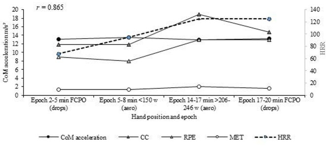
There were strong correlations between whole body CoM acceleration, MET and RPE (r = 0.865, p < 0.0001) that yielded the equation:
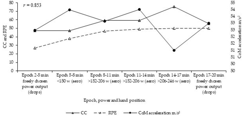
Replacing MET for caloric cost resulted in an equally strong correlation (r = 0.853, p < 0.0001) (Figure 3).
Discussion
This study investigated the relationship between whole body CoM acceleration, energy expenditure determined by the metabolic equivalent (MET), the rating of perceived of exertion (RPE) and heart rate reserve (HRR) during a typical triathlete indoor spin cycling training session. Power and the body position were varied to identify differences in acceleration patterns. The major experimental finding was that of extremely large effects pertaining to mediolateral acceleration during six minutes of steady-state cycling at 152-205 W as well as concurrent increases in longitudinal and anteroposterior CoM acceleration along with the RPE and the MET (Table 2). Additionally, switching from an aero to drops hand position induced extremely large effects in anteroposterior acceleration. Together, these combined variables may affect steady state triathlete spin cycling performance of longer duration.
Variations in mediolateral acceleration were most salient at epochs 8–11 min to 11–14 min which corresponded to constant power output of >152-205 W. Although this period of steady state cycling implies a sustainable, moderate effort which is below the lactate threshold (Ghosh, 2004), the effect of individual training status should be considered. In this instance, individual training volume and hours dedicated to cycling could have influenced results. It is also possible that gender-related differences in the pelvic tilt could arise, in part, from the bicycle set-up that was used. In particular, participants were asked to configure the handlebar and saddle position to their preferred settings, meaning that the change in the body position (i.e., from drops to aero) may not have been correctly configured, which may have required the female participant to rotate further forward when moving to the drops position. Along this line, gender-related differences in pelvic orientation when changing the body position have been observed with females exhibiting a greater anterior tilt in the drops position compared with males (Sauer et al., 2008). This could explain the high dispersion of standard deviation observed in the current study, noticeable in both longitudinal and anteroposterior magnitudes relative to increases in power. As the absolute magnitude of CoM acceleration would likely change between genders, further research is warranted to assess changes in performance settings.
In the current study, participants exhibited small effects in mediolateral acceleration when moving from the aero to drops hand position towards the end of the protocol. Regarding this change, as power went from high values of 206-246 W in aero cycling to freely chosen power in the drops, it is feasible that participants deliberately selected a lower power range as the protocol neared conclusion to anchor the perceptual exertion range (Figures 2 and 3). If, instead, the differences were attributable to anatomical factors, these did not seem to be captured by simple measures of acceleration magnitude. However, anteroposterior trunk acceleration could explain intraindividual differences in CoM motion because of the high magnitudes observed during the final minutes of cycling. This potentially represents the gross motion of the participant’s CoM around the anteroposterior axis. Equally, the possibility of poor trunk strength cannot be ruled out. A low level of core muscle strength can cause greater upper movement (Rannama et al., 2017). This would suggest a destabilising effect of the trunk and inefficient movement patterns leading to increases in whole body CoM acceleration.
The data in the current study suggest that triathletes increased whole body CoM acceleration magnitude during constant power as evidenced through minutes 8-11 to 14-17, respectively. The absolute increase in whole body CoM acceleration magnitudes combined with the RPE, the MET and HRR suggest an increase in participants’ work. Thus, the power transfer from the trunk to the lower body would increase with power due to an increase in hip reaction force, a result consistent with that found by others (Costes et al., 2015; Mestdagh, 1998). This warrants the need to quantify postural deviations to determine the influence of stability as it is currently unknown whether consistent trends would emerge for extended duration of spin cycling. Along this line, a structured strength and conditioning program with the focus on core trunk and hip strength may also reduce these effects.
In order to address whether whole body CoM acceleration was affected by changes in the body position and power, values were obtained at freely chosen power output at the commencement and conclusion of the protocol. When the stabilising role of the trunk is considered, reduced trunk lean can occur early as muscle fatigue can precede changes in trunk lean and trunk range of motion (Dingwell et al., 2008). Similarly, a significant compensatory effect on movement kinematics without alterations in pedalling would occur (Abt et al., 2007). Thus, it is possible that a similar attributable effect occurs through whole body acceleration of the CoM which may contribute to greater acceleration magnitudes and the RPE with a corresponding reduction in power.
The concomitant increases in the MET, RPE and HRR put increased emphasis on the blended physiological and temporal kinematics relationship. This is comparable to that reported by Zinoubi et al. (2018) and supports the premise that the RPE is associated with physiological variables (Kasimay Çakir et al., 2012). Although this increase was consistent until <206–246 W at which point the RPE levelled to a freely chosen cadence at minutes 17–20, an increase in the workload would theoretically lead to higher activation of the majority of muscles and therefore CoM trunk acceleration. As the RPE increases with fatigue (Rietjens et al., 2005), taken together with an increase in whole body CoM acceleration and power, it is plausible that excessive accelerations of the CoM could provide a technique to investigate postural changes during frequent spin cycle training sessions. Future research should determine variables which reflect muscular, whole body CoM acceleration and RPE status. Nevertheless, it is important to note that the RPE is affected not only by physical stress, but also psychological stress (Kumar et al., 2013), and therefore, the changes seen in HRR in the present study may also provide information not only on whether the athlete is physically able to train, but their mental readiness (i.e., holistic psychophysiological athlete preparedness). Furthermore, at submaximal levels exertion could be due to neurological alterations owing to reduced force despite increases in perception of effort (Noakes and St Clair Gibson, 2004). However, care must be taken when interpreting these results as data from the current study show the effect of a single cycling session with no direct measure of neuromuscular fatigue markers obtained.
This study provides information relevant for plausible associations between triathlete steady state spin cycling, whole body CoM acceleration, HRR, RPE and the MET. Although observable trunk movement can be indicative of an incorrect saddle position, it would be anticipated that increases in the RPE and the MET would ensue due to incremental increases in power. Notably, if participants increased power a concurrent increase in muscular force would occur which would conceivably increase the amount of mechanical work. Although insufficient to completely explain the increase in the RPE or to indicate its effects on whole body CoM acceleration, spin cycling performance and postural control, the use of musculoskeletal modelling to simulate pedalling and body acceleration during higher intensities may provide greater insights.
Whereas Galy et al. (2003) required cycling to fatigue, compared to the moderately short and non-fatiguing protocol used in this pilot study, it is challenging to draw conclusions. The use of steady state spin cycling negated participants performing a fatiguing bout of cycling, as might be experienced in a triathlon race. Whilst diligence must be applied to assess how kinematics might change with greater levels of intensity, this information, along with systematic evaluation of triaxial CoM acceleration distributions, could prove useful for training interventions.
Practical implications
The findings of this study have practical implications for coaches and triathletes. The quantification of energy expenditure based on accelerometric data in triathletes during a representative training session could be a useful and cost-effective metric to use in a real-time setting. This will ensure coaches and athletes can monitor and adjust whole body trunk CoM motion in accordance with the RPE, HRR and the MET efficiently throughout training.
Conclusions
An accelerometer, in combination with the RPE, the MET and HRR, may provide a practical, non-invasive, accessible and economically valid tool to monitor whole body CoM acceleration in triathletes. The use of a simple and highly accessible accelerometer tool means coaches and athletes can adopt the methods used within the study at minimal expense and without extensive training. This pilot study sets the foundation for further evaluation of a sensor-based approach to trunk CoM acceleration in triathlon cycling in order to take the measures reported here towards greater reductions in trunk accelerations.